Home
/ Arteries Diagram : Circle Of Willis Anatomy Function And What To Know - Anatomy and function of the coronary arteries.
Arteries Diagram : Circle Of Willis Anatomy Function And What To Know - Anatomy and function of the coronary arteries.
Arteries Diagram : Circle Of Willis Anatomy Function And What To Know - Anatomy and function of the coronary arteries.. Two major coronary arteries branch off from the aorta near the point where the aorta and the left ventricle meet. Like maps, the various diagrams emphasize different aspects. Blood vessels are the channels or conduits through which blood is distributed to body tissues. Create healthcare diagrams like this example called arteries and veins of the arm in minutes with smartdraw. Coronary arteries supply blood to the heart muscle.
Human body artery diagram in detail. To understand the system, the students need to create their diagrams. The abdominal aorta bifurcates at the level of the fourth lumbar vertebra to form the two common iliac arteries, each of which further branches into the external and the internal iliac artery. Inner body parts with their names. The defect is associated with narrowing of the trachea (windpipe) and bronchi (airways).
Arteries Of The Body Picture Anatomy Definition More from human.biodigital.com This is the opposite function of veins, which transport blood to the heart. Learn the differences between an artery and a vein. John bavosi/science photo library/getty images. Coronary arteries supply oxygenated blood to the heart muscle, and cardiac veins drain away the blood once it has been deoxygenated. Arteries of the head and neck diagram art print vintage anatomy art print on tea stained paper dog art dog s wfh office art. Inner body parts with their names. After receiving blood directly from the left ventricle of the heart, the. The tunica medica, which is the very muscular middle layer in arteries, is thinner and less muscular in veins.
Blood is pumped from the heart in the arteries.
To understand the system, the students need to create their diagrams. This is a congenital defect in which the left pulmonary artery branches off the right pulmonary artery, rather than directly from the pulmonary trunk. Arteries of the lower limb thigh leg foot the main artery of the lower limb is femoral artery it is a continuation of the external iliac artery terminal branch of the abdominal aorta the arteries and veins of the leg smartdraw arteries and veins of the leg create healthcare diagrams like this example called arteries and veins of the leg in minutes with smartdraw. Constricted arteries oppose blood flow, and more pressure is required to push blood. Some are more conceptual, others focus on branching, while still others attempt to preserve a spatial representation. The tunica medica, which is the very muscular middle layer in arteries, is thinner and less muscular in veins. Other arteries of the neck. Major arteries leading from the heart (via the ascending aorta) include the brachiocephalic artery, the left common carotid artery, and the left subclavian diagram 2(b) includes additional information about structures concerned with the system of electrical conduction operating in the heart, which is. Blood is transported in arteries, veins and capillaries. Arteries and veins of the arm. Arteries are components of the cardiovascular system. It can also help them in getting an overview of artery vs. The two exceptions are the pulmonary and the umbilical arteries, which carry deoxygenated blood to the organs that oxygenate it (lungs and placenta.
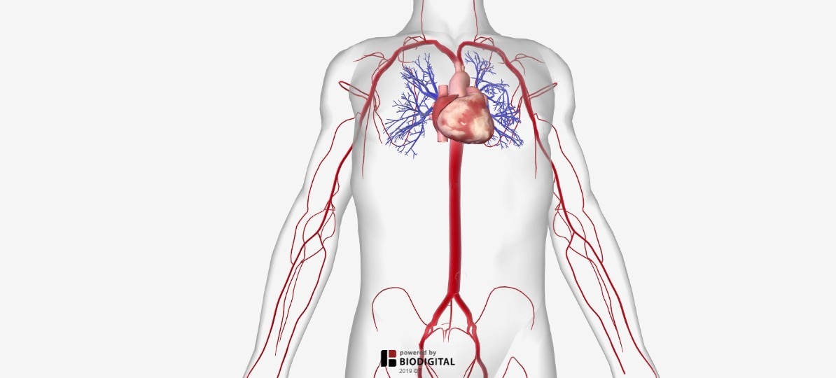
In this image, you will find external carotid artery, internal carotid artery, vertebral artery, aorta and arch, pulmonary artery, cardiac artery, thoracic aorta, celiac trunk, superior mesenteric artery, renal artery, gonadal artery, inferior mesenteric artery, common iliac artery, external iliac artery. 5 out of 5 stars. Create healthcare diagrams like this example called arteries and veins of the arm in minutes with smartdraw. Inner body parts with their names. Learn vocabulary, terms, and more with flashcards, games, and other study tools.
Basilar Artery Neuroangio Org from www.neuroangio.org Arteries and arterioles carry oxygenated blood _____ from the heart to the body. John bavosi/science photo library/getty images. The tunica medica, which is the very muscular middle layer in arteries, is thinner and less muscular in veins. Learn vocabulary, terms, and more with flashcards, games, and other study tools. Major arteries by definition, an artery is a vessel that conducts blood from the heart to the periphery. Coronary circulation is the circulation of blood in the blood vessels that supply the heart muscle (myocardium). This is a congenital defect in which the left pulmonary artery branches off the right pulmonary artery, rather than directly from the pulmonary trunk. 5 out of 5 stars.
The arteries' smaller branches are called arterioles and capillaries.
14+ heart arteries diagram labeled. 5 out of 5 stars. Arteries are components of the cardiovascular system. Arteries carry blood away from the heart in two distinct pathways: Resistance (r) the force opposing blood flow. Bodytomy provides a labeled iliac artery diagram to help you understand the anatomy and function of the common iliac. These arteries and their branches supply all parts of the heart muscle with blood. This is the opposite function of veins, which transport blood to the heart. The coronary arteries wrap around the outside of the heart. An artery (plural arteries) (from greek ἀρτηρία (artēríā) 'windpipe, artery') is a blood vessel that takes blood away from the heart to one or more parts of the body (tissues, lungs, brain etc.). To understand the system, the students need to create their diagrams. The right and left subclavian arteries give rise to the thyrocervical trunk. Arteries and veins of the arm.
Labeled heart diagram showing the heart from anterior. Because the rest of the body, and most especially the brain, needs a steady supply of oxygenated blood that is free of all but the slightest. The right and left subclavian arteries give rise to the thyrocervical trunk. Coronary circulation is the circulation of blood in the blood vessels that supply the heart muscle (myocardium). Blood is pumped from the heart in the arteries.
Arteries And Veins Blood Vessel Diagram The Circulatory System from sites.google.com It can also help them in getting an overview of artery vs. The narrowed arteries are at higher risk for complete blockage from a sudden. This process is called atherosclerosis. Over the years, cholesterol plaques can narrow the arteries supplying blood to the heart. pulmonary artery sling can be treated surgically. 14+ heart arteries diagram labeled. John bavosi/science photo library/getty images. Only 3 available and it's in 2 people's carts.
This process is called atherosclerosis.
After receiving blood directly from the left ventricle of the heart, the. It is returned to the heart in the veins. Learn vocabulary, terms, and more with flashcards, games, and other study tools. These vessels are channels that distribute blood to the body. Bodytomy provides a labeled iliac artery diagram to help you understand the anatomy and function of the common iliac. The cardiovascular system consists of the heart, blood vessels, and the approximately 5 liters of blood that the blood vessels transport. Smartdraw includes 1000s of professional healthcare and anatomy chart templates that you can modify and make your own. The veins also lack the elastic internal lamina that lies. In this image, you will find external carotid artery, internal carotid artery, vertebral artery, aorta and arch, pulmonary artery, cardiac artery, thoracic aorta, celiac trunk, superior mesenteric artery, renal artery, gonadal artery, inferior mesenteric artery, common iliac artery, external iliac artery. Blood vessels are the channels or conduits through which blood is distributed to body tissues. The vessels make up two closed systems of tubes that begin and end at the heart.one system, the pulmonary vessels, transports blood from the right ventricle to the lungs and back to the left atrium.the other system, the systemic vessels, carries blood from. Learn the differences between an artery and a vein. This is a congenital defect in which the left pulmonary artery branches off the right pulmonary artery, rather than directly from the pulmonary trunk.